Genomics of Giant Bacteria Found in Guadeloupe Mangroves
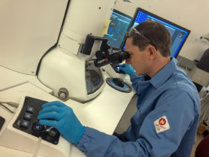
Jean-Marie Volland at Donner lab. (Massie S. Ballon)
In Science, a team led by Jean-Marie Volland, a scientist with joint appointments at the Department of Energy (DOE) Joint Genome Institute (JGI) and the Laboratory for Research in Complex Systems, and Silvina Gonzalez-Rizzo and Olivier Gros of the Université des Antilles, described the morphological and genomic features of a giant filamentous bacterium, along with its life cycle.
Volland got involved with the giant Thiomargarita bacteria when he returned to the Gros lab as a postdoctoral fellow. When he applied to the discovery-based position at the LRC that would see him working at the JGI, Gros allowed him to continue research on the project. Volland began studying Ca. T. magnifica in Tanja Woyke’s Single Cells Group to better understand what this sulfur-oxidizing, carbon fixing bacterium was doing in the mangroves. “We started this project under the JGI’s strategic thrust of inter-organismal interactions, because large sulfur bacteria have been shown to be hot spots for symbionts,” said Woyke, who also heads the JGI’s Microbial Program and is one of the article’s senior authors. “Yet the project took us into a very different direction.”
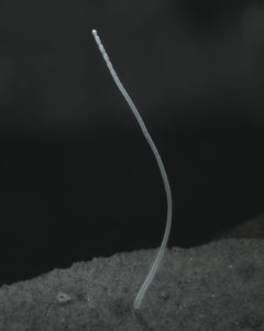
Single filament of Ca. Thiomargarita magnifica (Jean-Marie Volland)
The JGI team then used single-cell genomic sequencing to analyze five of the bacterial cells on the molecular level. Sequencing reads from these cells were assembled and binned into genomes, then checked for quality and assessed for taxonomy using apps in the KBase platform. Using imaging facilities available at Berkeley Lab, such as confocal laser scanning microscopy and transmission electron microscopy (TEM), allowed Volland to visualize the filaments and the cell membranes in more details. These techniques allowed him to observe novel, membrane-bound compartments that contain DNA clusters. He dubbed these organelles “pepins,” after the small seeds in fruits.
Other JGI and Biosciences authors on the paper are Tomáš Tyml, Natalia Ivanova, Frederik Schulz, Danielle Goudeau, Nathalie H. Elisabeth, Nandita Nath, Daniel Udwary, Rex R. Malmstrom, Karen M. Davies, and Nigel J. Mouncey.
Learn more on the JGI website.
Publication: J.M.* Volland, S.* Gonzalez-Rizzo, O.* Gros et al. “A centimeter-long bacterium with DNA contained in metabolically active membrane-bound organelles.” Science. (2022). [doi: 10.1126/science.abb3634]
*co-first authors